ZEISS Sigma 高画質イメージングと高度な解析を実現する電界放出型SEM
ZEISS Sigmaファミリーは、電界放出型走査電子顕微鏡(FE-SEM)のテクノロジーと優れたユーザーエクスペリエンスを兼ね備えています。イメージングと解析ルーチンを構造化することで生産性が向上し、新しい材料、品質検査用の粒子試料、生物・地質試料などの観察に活用できます。妥協のない高分解能イメージングにより、1 kV以下の低加速電圧でも高い解像度とコントラストを達成します。さらに、クラス最高のEDSジオメトリを使用して高度な顕微鏡解析を実行し、より正確な解析データを2倍の速さで取得することができます。
Sigmaファミリーは、ユーザーを高度なナノ解析の世界へと導きます。


Sigma 360
コア施設から選ばれる直感的なイメージング
- セットアップからAIによる結果取得まで、専用のガイドに従って操作するだけの直感的なイメージングワークフローをご体験ください。
- 1 kV以下でも優れた解像度と最適なコントラストを実現
- VPイメージングにより、非導電体の非常に困難な条件でも優れた結果を取得可能
キャプション:NanoVP liteモードでイメージングしたポリスチレン。
-


直感的なイメージングワークフロー
セットアップからAIによる結果取得まで
- 初心者でも専門知識に基づいた結果取得が可能です。使いやすく覚えやすいワークフローにより、ナビゲーションから後処理までの各ステップが効率化され、高速イメージングを実現します。また、トレーニングに必要な時間も短縮できます。
- ZEISS SmartSEM Touchのソフトウェア自動化によって、ナビゲーション、パラメータ設定、画像取得を開始できます。
- ここで、タスク固有のツールキットを備えた、後処理に最適なZEN coreが役立ちます。最も推奨されているのは、機械学習に基づく画像のセグメンテーションが可能なAI Toolkitです。Connect Toolkitではマルチモーダル実験を組み合わせられます。また、Materials Appsによる微細構造、粒径、層厚の解析も可能です。
-

1 kV以下で強みを発揮
優れた解像度と最適なコントラスト
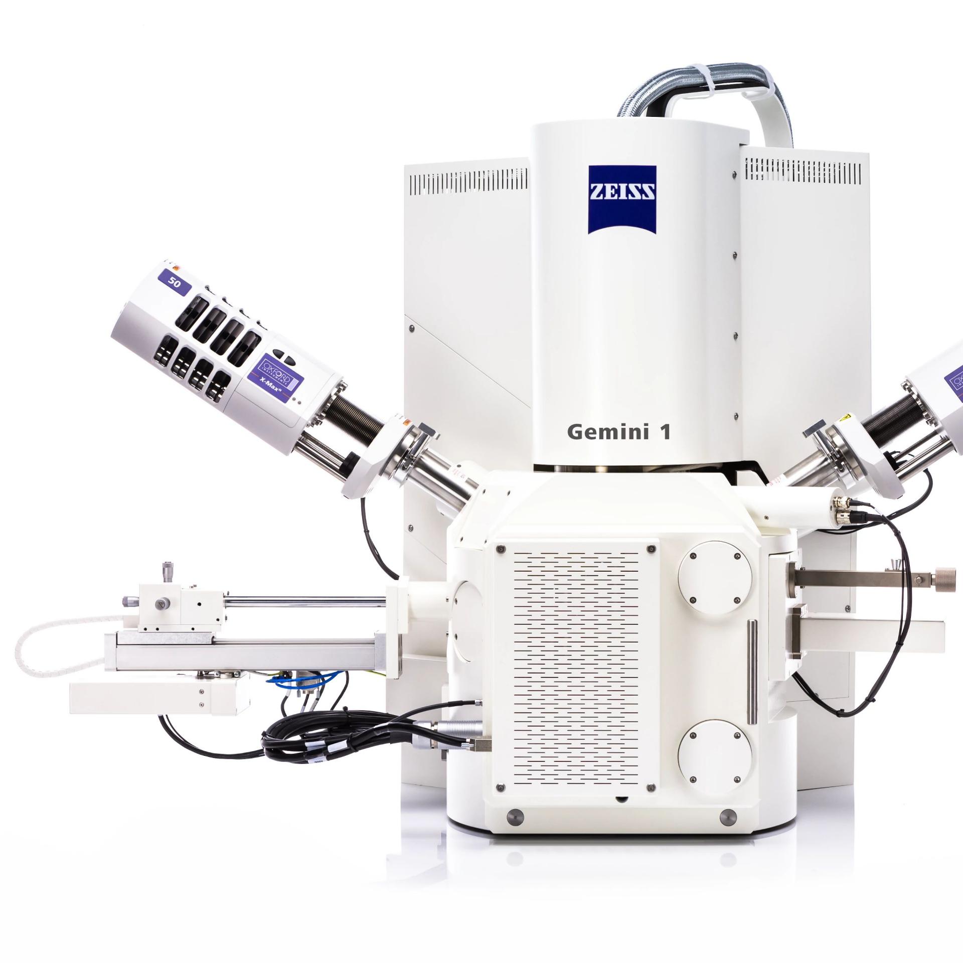
- イメージング・解析パフォーマンスの鍵となるのは光学カラムです。ZEISS Gemini 1電子光学系を搭載したSigmaは、特に低加速電圧において、あらゆる試料に対して優れた分解能を発揮します。
- Sigma 360の低電圧分解能の仕様は、1.9 nmで500 Vになりました。色収差を最小限に抑えることで、1 kVの分解能が1.3 nmで10%以上向上しています。
- 低真空モードでの後方散乱検出や観察が困難な試料でも、これまで以上にイメージングが容易になりました。
-

非常に困難な条件でもVPイメージングを実現
解析・イメージング用NanoVP liteモード
- 新登場のNanoVP liteモードと新しい検出器が、5 kV以下における非導電体からの高品質データの取得を簡素化します。
- 強化されたイメージングとEDS解析により取得時間が短縮され、表面感度の高い情報と、高速EDSマッピングのための一次ビーム電流が提供されます。
- aBSD1(環状後方散乱電子検出器)や次世代C2D(カスケード電流検出器)などの新しい検出器を使用することで、低加速電圧でも優れたイメージングが可能です。


ナノスケール球体の段丘の測定サイズは3 nm。Sigma 560、Inlens SE検出器、500 Vで取得。")
ナノスケール球体の段丘の測定サイズは3 nm。Sigma 560、Inlens SE検出器、500 Vで取得。")
Sigma 560
ハイスループット解析とin situ実験の自動化
- リアルワールドの試料を効率的に解析。スピードと汎用性を兼ね備えたSEMベースの解析が可能です。
- In situ実験の自動化:無人検査のための完全統合型ラボを実現します。
- 1 kV以下のイメージングが困難な試料にも対応。包括的な試料情報を収集できます。
-

リアルワールドの試料を効率的に解析
EDSで汎用性の高い観察と高速イメージングを実現
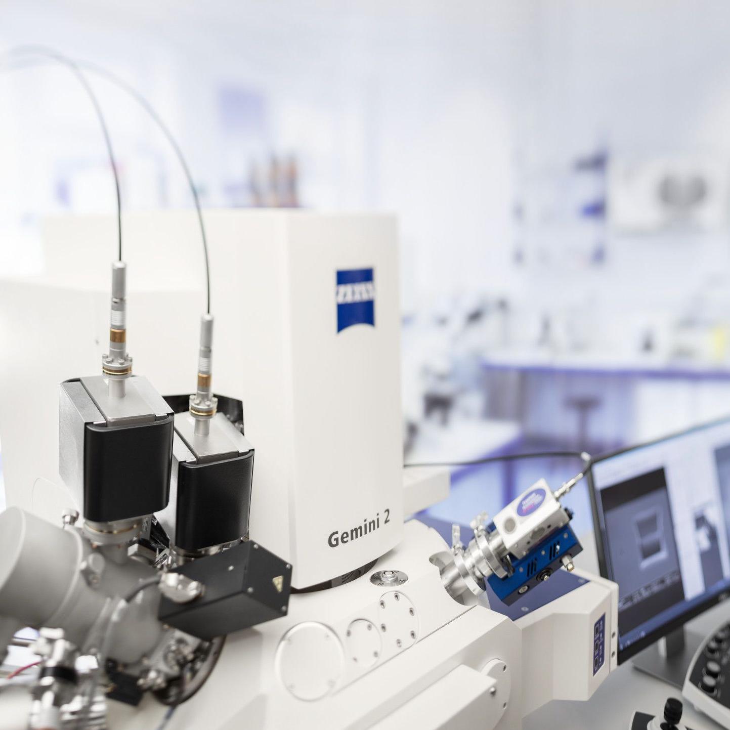
- Sigma 560のクラス最高のEDSジオメトリにより、解析の生産性が向上します。180°正反対に位置する2つのEDSポートが、低ビーム電流や低加速電圧においても優れたスループットとシャドーフリーマッピングを保証します。
- EBSDとWDS用のポートをチャンバーに追加することで、EDSをさらに上回る解析が可能になります。
- 新しいNanoVP liteモードにより、非導電体に対しても高いシグナルとコントラストで解析が可能です。
- 新開発のaBSD4検出器なら、高度なトポグラフィーの試料でも容易にイメージングできます。
- Sigma 560のクラス最高のEDSジオメトリにより、解析の生産性が向上します。180°正反対に位置する2つのEDSポートが、低ビーム電流や低加速電圧においても優れたスループットとシャドーフリーマッピングを保証します。
-

In situ実験の自動化
無人検査のための完全統合型ラボを実現
- 完全統合型ソリューションであるSigma向けin situラボは、自動化されたワークフローで、無人の加熱・引張試験の結果を取得します。
- 3DのSTEMトモグラフィーやAIベースの画像セグメンテーションなど、ナノスケールの3D解析によってワークフローをさらに拡張可能です。
- 新開発のaBSD4では、3Dサーフェスモデリング(3DSM)をライブで実行できます。
-

イメージングが困難な試料にも対応
1 kV以下で強みを発揮
- 1 kV、500 Vでも豊富な情報量のイメージング・解析を実現:Sigma 560の低電圧分解能の仕様は500 Vで1.5 nmです。
- 新しいNanoVP liteモードでは、低真空などの困難な条件でも新たに搭載されたaBSDまたはC2D検出器を使用することで、最低3 kVの低加速電圧で観察が可能です。
- 電子機器の調査をするには、クリーンな環境を維持する必要があります。プラズマクリーナー(推奨)や、6インチウェーハのシャトル化を可能にする大型エアロックを仕様することで、チャンバーを不純物による汚染から保護できます。


テクノロジー


ビームブースター、Inlens検出器、Gemini対物レンズを搭載したGemini光学カラムの断面図。
Gemini 1光学系
Gemini 1の光学系は、対物レンズ、ビームブースター、Inlens検出コンセプトの3つの要素で構成されています。対物レンズの設計には静電場と磁場が組み合わせられており、磁場が試料に与える影響を最小限に抑えながら、光学性能を最大限に高めます。これにより、磁性材料のように困難な試料であっても優れたイメージングが可能となります。Inlensは、二次電子(SE)や後方散乱電子(BSE)を検出することにより、イメージング時間を最大限短縮しつつ、効率的なシグナル検出を実現することをコンセプトにしています。ビームブースターテクノロジーが小さなプローブサイズと高いSN比を保証します。


Gemini 1の光学カラムと検出器の断面図。
検出器搭載SigmaのGemini 1のカラム。1 Inlens検出器(SEまたはDuo)。2 ETSE検出器、3 VPSE、4 C2D、5 aSTEM、6/7 EDS検出力が高く、様々な後方散乱を感知する検出器(aBSD1など)。
柔軟な検出機能
様々な検出器を搭載したSigmaでは、最新の検出テクノロジーにより、あらゆる試料の特性評価が可能です。ETSEと高真空モード用のInlens検出器が、高分解能のトポグラフィー取得をサポートします。低真空モードでVPSEやC2D検出器を使用することで、鮮明な画像を取得できます。aSTEM検出器では高分解能の透過画像を生成できます。様々なオプションが揃ったBSE検出器(aBSD検出器など)で組成やトポグラフィーを観察できます。
、電子ビームのスカート(緑)。")
、電子ビームのスカート(緑)。")


NanoVP liteモード
- NanoVP liteモードでは、スカート効果が低減され、ビームガスの光路長(BGPL)が短くなります。スカートを抑えることで、SEおよびBSEイメージングにおけるSN比が向上します。
- 5つの環状セグメントを持つ格納式のaBSDは、優れた材料コントラストを実現します。ビームスリーブを搭載しており、NanoVP liteモードでの操作時はポールピースの下に設置されます。ハイスループットと低加速電圧での組成・トポグラフィーコントラストイメージングを実現し、VPやHV(高真空)に適しています。


")
")
")
")


")
")
")
")


またはガーネット含有片麻岩。")
またはガーネット含有片麻岩。")
またはガーネット含有片麻岩。")
またはガーネット含有片麻岩。")


アクセサリ


ZEISS FE-SEM用In Situラボ
材料性能を微細構造に関連付ける
ZEISS FE-SEMに、加熱・引張実験用のin situソリューションを追加できます。統合ソリューションのメリットをご活用ください。金属、合金、ポリマー、プラスチック、複合材料、セラミックスなどの材料を観察可能です。さらに、機械的引張または圧縮ステージ、加熱ユニット、専用高温検出器を解析と組み合わせることができます。統一されたソフトウェア環境が無人自動材料試験を可能にし、1台のパソコンからすべてのシステムを制御できます。


SmartEDX
組み込み式エネルギー分散型X線分光法


完全統合型RISE
ラマンイメージングと査型電子顕微鏡のメリットを活用
ラマン分光イメージング(RISE)を追加すれば、材料の特性評価を補完できます。Sigma 360を共焦点ラマンイメージング機能で拡張することで、試料の化学的フィンガープリントの取得が可能になります。分子・結晶の情報を認識することができます。また、ラマンマッピングとEDSデータを使用して、3D解析およびSEMイメージングとの相関が可能です。完全統合型RISEにより、クラス最高のSEMおよびラマンシステムの両方を活用できます。
関連アプリケーション
ダウンロード
-
-

3D Imaging Systems
Your Guide to the Widest Selection of Optical Sectioning, Electron Microscopy and X-ray Microscopy Techniques.
ファイルサイズ: 5 MB -

ZEISS Sense BSD
高速で試料ダメージが少ない微細構造 イメージングのための後方散乱電子検出器
ファイルサイズ: 13 MB -

ZEISS Sigma 300 with RISE
Extend your ZEISS Sigma 300 with Fully Integrated Raman Imaging and Scanning Electron Microscopy (RISE)
ファイルサイズ: 2 MB -

ZEISS Sigma Family
高画質イメージングと高度な分析のための 電界放出型走査電子顕微鏡(FE-SEM)
ファイルサイズ: 42 MB -

ZEISS SmartEDX
The ZEISS Embedded EDS Solution for Your Routine SEM Microanalysis Applications
ファイルサイズ: 2 MB -

工業用セラミックス研究のためのZEISS顕微鏡ソリューション
先端セラミックス設計のための2D、3D、4Dソリューション
ファイルサイズ: 1 MB -

Reduced Energy Consumption
Optimized Operating Efficiency
ファイルサイズ: 340 KB -

ZEISS FE-SEM用in situラボ
材料の性能を微細構造と関連付ける
ファイルサイズ: 4 MB -

ZEISS Sigma Family - Flyer
Your FE-SEMs for High Quality Imaging & Advanced Analytical Microscopy
ファイルサイズ: 2 MB
-
-
-

Large Volume Imaging of Eye Muscle by SIGMA VP and 3View
Serial Block Face Imaging
ファイルサイズ: 1 MB -

ZEISS Sigma 300 with WITec Confocal Raman Imaging
Characterizing Structural and Electronic Properties of 2D Materials Using RISE Correlative Microscopy
ファイルサイズ: 6 MB -

Voltage Contrast in Microelectronic Engineering
ファイルサイズ: 1 MB -

ZEISS LaserSEM
Your solution for site-specific preparation from the meso- to the microscale – a femtosecond laser integrated into a ZEISS FE-SEM
ファイルサイズ: 2 MB -

Case Study
Corrosion analysis of modern and historic railway trackwith optical, electron and correlative Raman microscopy
ファイルサイズ: 7 MB -

Cathodoluminescence of Geological Samples: Fluorite Veins
ZEISS Scanning Electron Microscopes with Atlas
ファイルサイズ: 5 MB -

Investigating Sweet Spot Imaging of Perovskite Catalysts Bearing Exsolved Active Nanoparticles
ファイルサイズ: 5 MB -

The building blocks of our solar system
Studying the Winchcombe meteorite
ファイルサイズ: 3 MB -

ZEISS Microscopy Solutions for Geoscience
Understanding the fundamental processes that shape the universe expressed at the smallest of scales
ファイルサイズ: 15 MB -

ZEISS Microscopy Solutions for Oil & Gas
Understanding reservoir behavior with pore scale analysis
ファイルサイズ: 7 MB -

ZEISS Sigma 300
Quantitative EBSD Studies of Soft Magnetic Composites
ファイルサイズ: 10 MB
-